The revolutionary mammogram technique that may be just too revolutionary


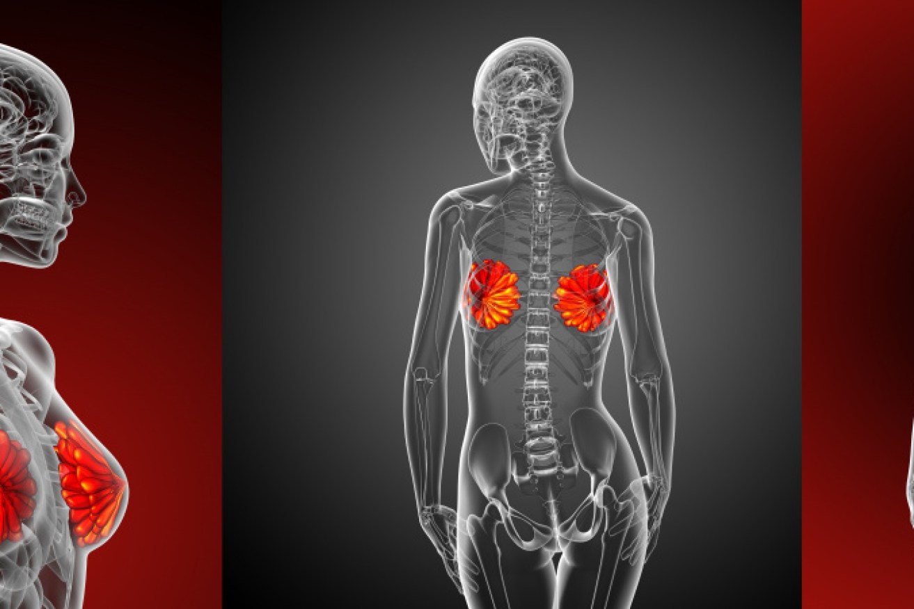
Many women fighting breast cancer will soon see the cost of a key medication drastically reduced. Photo: Getty
Sydney researchers have conducted the first Australian population-based trial of a 3D mammography technique and found that it detects significantly more cancers than standard mammography.
A US study last week came to the same conclusion: digital breast tomosynthesis results in an overall increase in cancer detection rates “irrespective of the tumour type, size, or grade of cancer”.
Overseas the technique is being hailed the next big thing. A 2017 article in the American Journal of Roentology predicted the screening technique will likely replace the conventional approach we now know.
But not yet.
BreastScreen Australia, the federal government’s population-based screening program for women, says that “robust evidence is required before tomosynthesis could be used as a routine screening tool” – and that its use should be “be confined to clinical trial settings”. (You can read BreastScreen’s full position statement here.)
The known problems with tomosynthesis: the gains in cancer detection may be offset by larger radiation doses, longer screen-reading times, plus false positives. And it remains an open question as to whether the technique – increasingly available in a clinical setting – will lead to better health outcomes.
So what is digital breast tomosynthesis?
Nehmat Houssami is Professor of Public Health and National Breast Cancer Foundation Research Leadership Fellow at the University of Sydney’s Faculty of Medicine and Health. She led the Australian study.

Leader of the study: Nehmat Houssami, Professor of Public Health and National Breast Cancer Foundation Research Leadership Fellow at the University of Sydney’s Faculty of Medicine and Health. Photo: University of Sydney
In an email interview, she explained the new 3D technique:
“3D mammography technology (breast tomosynthesis), is similar to standard (‘regular’ or 2D) mammography in that it takes an X-ray of the breast while it is compressed (for a short time).
“When the test is done, the main difference is that compared to 2D mammography, the 3D technology takes several small dose X-rays at various angles and then recreates a ‘near 3D’ image of the breast.
“What that achieves: it reduces some of the overlapping (superimposed) breast tissue that can cause problems on mammograms. By problems I mean that overlapping tissue layers can hide a small cancer or can create ‘lesions’ from normal tissue structures.
“So, 3D technology can help improve interpretation of the images by ‘spreading out’ the tissue layers of the breast: that tends to improve cancer detection or it can reduce false-positive screening results (‘false alarms’).
“But we should be aware that the findings on this technology have varied between studies. That is why it is important to carry out our own studies here in Australia.”
The Australian study
The researchers compared tomosynthesis and standard mammography screening of women attending Maroondah BreastScreen in Victoria, for routine breast screening between August 18, 2017, and November 8 last year.
Thousands of screenings were undertaken: 5018 tomosynthesis and 5166 standard mammography screens.
They found:
- Tomosynthesis detected 49 cancers (40 invasive, nine in situ). Standard mammography detected 34 cancers (30 invasive, four in situ). In situ refers to cancer in which abnormal cells have not spread beyond where they first formed.
- The estimated difference was 3.2 more detections per 1000 screens with tomosynthesis. The difference was greater for repeat screens and for women aged 60-plus.
- The recall rate was greater for tomosynthesis (4.2 per cent) than standard mammography (3 per cent). Recall rate refers to the percentage of screening mammograms that are interpreted as suspicious and require some sort of diagnostic follow-up imaging and/or biopsy.
- The median screen reading time for tomosynthesis was 67 seconds, compared to standard mammography (16 seconds).
The researchers concluded
Professor Houssami said: “The pilot trial has shown that 3D performs better than 2D as far as breast cancer detection (which is the good news) but there were some negatives as well – 3D technology causes more false-positive results and it increased the time to read the screen, it also meant there was a little more radiation to the breast.
These are the issues that need to be considered (all together) to decide the next step for using 3D in BreastScreen.”
She said that in many radiology practices, 3D mammograms are replacing the regular mammograms.
“But when it comes to population screening, very careful study of whether the benefits outweigh the harms from screening is necessary,” she said.
“Because population screening is about screening women without symptoms, we need to be sure there is a net benefit (improved health) from advocating a screening test or changing the existing screening test.
“While we have evidence on how 3D ‘detects’ we do not yet have evidence on the longer term health outcomes for 3D versus 2D screening, and this will be very important to do in future research.”
Professor Houssami said there is ongoing discussion with the national screening program about planning a larger study in Australia using the 3D technology “to help us gather the knowledge needed about whether and how the 3D mammogram (compared to 2D) affects outcomes for women”.
The results of the pilot study were published in October in the Medical Journal of Australia.


